

Predicting CT dosimetry in children via whole body mappings
Triangulated surfaces from an example deformed adult template and target child.
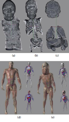
Estimating radiation dose accumulated by a patient throughout life is an important matter that has been receiving much attention recently, in particular for children (for example in the New England Journal of Medicine's recent critique of CT use and the adoption of the Image Gently program by the American Association of Physicists in Medicine). While directly measuring dose to individual organs is impractical, computational phantoms containing dosimetric information have been developed serving this purpose, such as the XCAT phantom. A key shortcoming of this strategy is that standard phantoms cannot adequately reflect variability between patients, particularly for children of different sizes and ages. The goal is to map fine structures in the adult XCAT phantom to several bodies of different sizes and ages. Here it is necessary to develop multi-kernel multi-manifold multi-modality diffeomorphic mapping methods that can faithfully map these fine structures to the pediatric targets. Currently we manually segmenting a subset of organs from pediatric CT data, using a scaled and well matching XCAT phantom to insert missing anatomy if necessary (for example the head, in a body scan), and calculating a mapping to a similarly segmented adult XCAT phantom. The resulting transformation is used to map rich anatomical information to the child's body.
Click Here for more about Dr. Ratnanather's work >>
View the research article "Patient Specific Dosimetry Phantoms Using Multichannel LDDMM of the Whole Body", Here >>
View the research article "Effects of protocol and obesity on dose conversion factors in adult body CT", Here >>